

HIND LIMB RUDIMENTS FOUND ON MODERN DAY WHALES
EXAMPLE #3
1956 -- Female Sperm Whale caught by Japanese whaling operation, Nov. 8th, 1956, “protuberances on both sides of the genital opening. … The height of the protuberance was 5.35 centimeters on the right side, 6.56 centimeters on the left side.” (SEE PHOTOS OF THE PROTRUSIONS FROM SIX DIFFERENT ANGLES, Fig. 5)
“Upon examining the interior of the left limb three partially cartilaginous bones were found. They correspond to pelvis, femur, and possibly to tibia, but no joints exist between them. Pretty strong muscles connect between femur and tibia. The tibia is 13 centimeters long for the greater part cartilaginous, and only partly ossified stick-like body with its distal end inserted into the skin of the hind-limb protuberance. (SEE THE SCHEMATIC DIAGRAM BASED ON THE DISSECTION [Fig. 6] AND ALSO SEE THE PHOTOGRAPH OF THE DISSECTED HIND LIMB RUDIMENTS [Fig. 7]) A number of arteries and nerves run parallel to this tibia distalward. … The veins are not easily visible by the naked eye, but they are found attached intimately to the wall of arteries. This case can be understood by assuming abnormal retention of the early embryonic state, and show very probably an atavism back to the quadripedal condition of the whale’s remote ancestors. It can never be a malformation of no phylogenetic significance.” “We searched into the interior of the left limb. The pelvic bone was found there. … In the neighborhood of the pelvis, nearly at the middle part of this bone, the femur covered with cartilage is present taking the form of a small ball with the diameter ca. 3 centimeters. … 4.8 centimeters distant from the femur a mostly cartilaginous stick of the length 13 centimeters is present. It is only partially ossified. ... It is difficult to determine whether this stick corresponds either to tibia, fibula, or both of them fused together, or rather to an isolated distal portion of femur. But we take it provisionally for tibia in view of two slender muscles coming from the femur, and inserting to the anterior surface of the bony part of the stick. … The distal end [of the tibia] lies in the central part of the hind limb protrusion.” (SEE THE SCHEMATIC DIAGRAM BASED ON THE DISSECTION [Fig. 6] AND ALSO SEE THE PHOTOGRAPH OF THE DISSECTED HIND LIMB RUDIMENTS [Fig. 7]) “Two weak muscles (M5 and M6 in Fig. 6) are attached to the osseous tibia by intercalation of tendons. For the time being we take these muscles for the rudimentary mm. vast. … Our attention was further given to the richness of nerves and arteries pertaining to the limb. All of them run nearly parallel to the tibia in the proximal-distal direction … As to large arteries we have counted six of them (A1, A2, A3, A4, A5, A6 in Fig. 6) at the proximal end of the tibia. It is noteworthy that most of them reach the interior of the protruded limb. … All of the nerves destined to the hind limb are continuous from a thick trunk (N1+2) passing through the triangular space between pelvis and femur mentioned above (S). … Nerves and arteries run at first ventral to the pelvis, then dorsal to the femur, to reach further the tibial region.”
SOURCE: Ogawa, R., and Kamiya, T. A. (1957) "Case of the Cachalot [Sperm Whale] With Protruded Rudimentary Hind Limbs." Scientific Reports of the Whales Research Insititute, No. 12, p. 197-208.
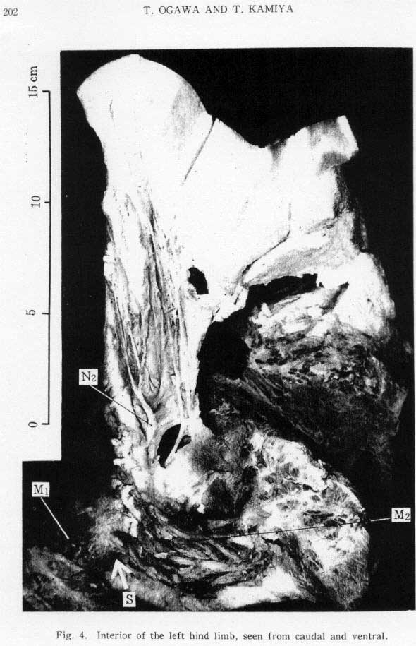
Figure 7
This photo goes with the previous example, EXAMPLE #3
No comments:
Post a Comment