HIND LIMB RUDIMENTS FOUND ON MODERN DAY WHALES
EXAMPLE #2
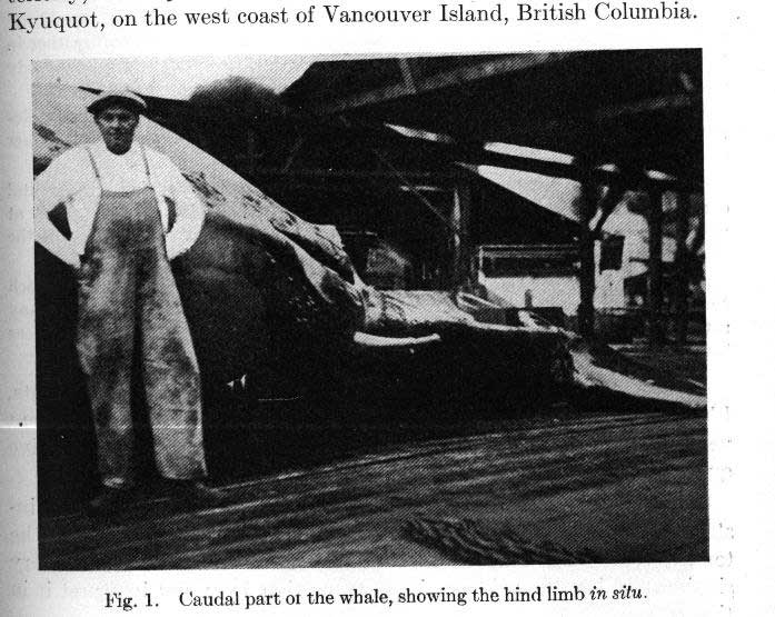
In July 1919, a female Humpback Whale with two remarkable protrusions on the ventral side of the body, posteriorly, was captured by a ship operating from the whaling station at Kyuquot, on the west coast of Vancouver Island, British Columbia [Canada]. One of the protrusions was cut off by the crew of the vessel but the other was photographed in situ by the superintendent of the Station. (Figure #3) Mr. Sidney Ruck and Mr. Lawson, officials of the Consolidated Whaling Company, appreciated the importance of the discovery and presented the skeletal remains of the attachment to the Provincial Museum, Victoria, B.C. [Canada]. At my request, Mr. Francis Kermode, Director of the Provincial Museum, very courteously submitted the bones to me with permission to publish upon the result of my examination.
Under date of March 4, 1920, Mr. Ruck writes to Kermode as follows: “I enclose herewith three photographs showing the unusual development of the pelvic Rudiments in a whale captured at the Kyuquot Station last July, of which you have the bones. It is to be regretted that better pictures in evidence of this unprecedented development were not obtained. I have been connected with the Whaling Industry for 22 years and during my time have come in contact with prominent Naturalists such a Professor True of the Smithsonian Institute, Professor Lucas of the Natural History Museum, New York, and neither in their experience or mine have the protrusion of the pelvic bones beyond the body ever been seen or heard of. This particular whale was a female Humpback of the average length with elementary legs protruding from the body about 4 feet 2 inches, covered with blubber about one-half an inch thick. As shown in the best photograph these legs protruded on either side of the genital opening; the left leg was cut off by the crew of the vessel and lost, and the point at which it was cut off is clearly shown in the photograph. The end of the leg seen in the picture terminated in a kind of round know like a man’s clenched fist. The two bones of the leg which you have are connected by cartilage which I was informed had shrunk about 10 inches, and possibly more by this time. At any rate the total length of the leg before it was cleaned of the blubber and flesh was, as before stated, about 4 feet, 2 inches, from the body.”…

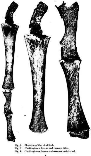
The skeletal remains in my possession consist of two bones and two heavy cartilages. When placed in position (see Figure #4), the total length is 31 inches.

FEMUR (a long cartilage) -- The larger bone is deeply concave proximally and to it is attached a massive cartilage which, in its present shrunken condition, is 5 ¼ inches in length and 1 5/8 inches wide. I estimate that this cartilage was at least 15 inches long and 3 inches wide when fresh. I believe that this cartilage represents the femur. It probably lay entirely within the body, its proximal end being attached to the pelvic vestiges. Such a massive cartilage much necessarily have had a firm support and leads me to believe that the pelvic elements in this individual were of extraordinary size. …
TIBIA (the longer of the two bones) -- The larger of the two bones I identify as the tibia. It is 14 ¼ inches in greatest length, is well developed, and has a hard, smooth outer surface. At the proximal end its greatest width is 3 ¾ inches, it narrows gradually for three-fourths of its length, and then suddenly expands at the distal extremity, where it is 2 ½ inches wide.
TARSUS (another cartilage) -- The distal end of the tibia is convex and gives attachment to a cartilage which in its shrunken state is 4 ¾ inches long and 1 ¾ inches wide. This cartilage, I believe, represents the tarsus. That it presents not ossifications is by no mean surprising as the carpal bones in the forelimbs of cetaceans are sometimes entirely absent and often in a more or less rudimentary condition. Mr. Ruck says “the two bones of the leg which you have are connected by cartilage which I was informed had shrunk about 10 inches and possibly more by this time.” This would give the tarsal cartilage a lengthy of nearly 15 inches. METATARSAL (the shorter of the two bones) -- The distal element in the leg is a hard, well-developed bone which I identify as a metatarsal. It has the characteristic shape of the metacarpals in the fore limbs of cetaceans except that it is more slender. It is 6 1/8 inches long. 1 7/8 inches in distal width; its least width is 15/16 of an inch. To the distal end of the metatarsal is attached a heavy cartilage of which only 3/4/ of an inch remains intact. This cartilage probably formed the extremity of the hind limb skeleton.
EXTERNAL APPEARANCE OF THE LIMB -- In reference to the limb as it appeared in the fresh condition, Mr. Ruck says that the end terminated in a “kind of round knob like a man’s clenched fist,” that the total length was about four feet and two inches, and that it was covered with blubber about one-half inch thick. I infer from Mr. Ruck’s description that the connective tissue and blubber were essentially the same as in the flipper, or fore limb, of cetaceans. The photograph of the limb in situ show that there are two prominent, truncated tuberosities on the distal half. The proximal “bunch” evidently indicates the distal end of the tibia and the other is at the extremity of the metatarsal. These tuberosities may very properly be homologized with those on the other, or anterior, edge of the flipper in the Megaptera (Humpback Whale) which indicate the extremities of the radius and the second digit. This is, I believe, a point which has considerable significance.

Figure 5
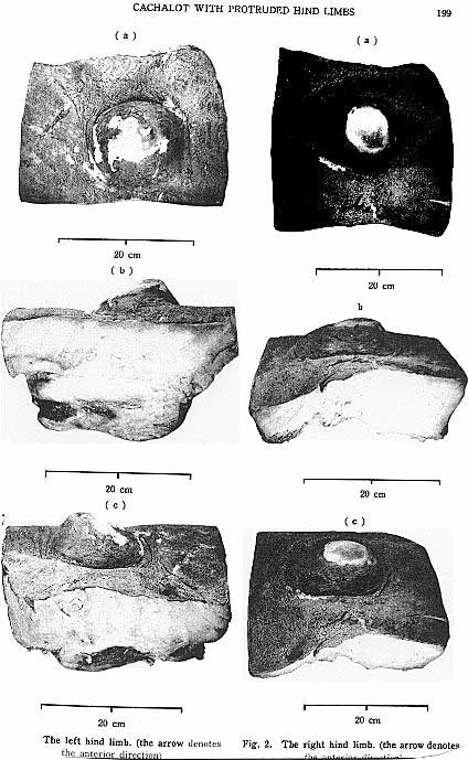
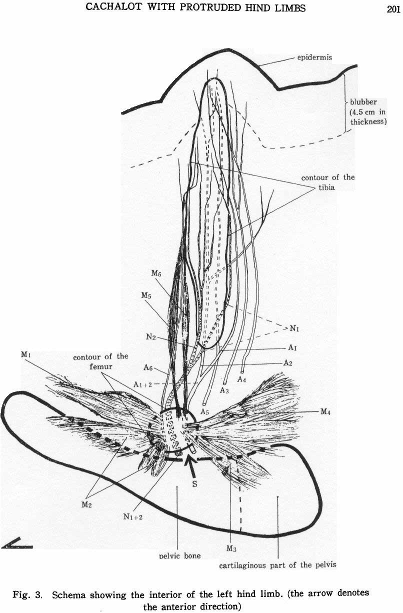
This photo goes with the following example, EXAMPLE #3
Since the stalk-like cartilaginous femur probably lay entirely within the body and the remainder of the limb entirely outside, there was undoubtedly a certain flexibility at the point of junction with the body.
In 1914 Professor W. Kikenthal described external rudimentary hind limbs in three early embryos of Megaptera (Humpback Whale). These appear as two more or less caudally directed papillae on either side of the genital organ in the same relative position as the himb limbs which I have described in this paper. In Kukenthal’s Stage 1 (an embryo 32 mm. In length) the rudiments are best developed and are 1.2 mm. Long. In Stage II the rudiments are somewhat less distinct…In Stage III (an embryo 30 mm. Long) the hind-limb rudiments have still more decreased in size and appear as minute papillae. Kukenthal and Guldberg have also discovered hind-limb rudiments in embryos of other cetacean species. Since Kukenthal’s and Guldenberg’s researches have shown that external hind-limb rudiments are still present in some cases [actually in “all” cases -- E.T.B.] in embryonic life, it is by no means impossible that, these vestigial organs should continue their growth and persist until the adult stage. I believe that that is exactly what has occurred in the specimen which I have described above, and that we are confronted with a clear case of partial reversion to a primitive quadripedal condition. The limbs, according to the statements of the whalers, were symmetrical; they are in the exact position in which hind-limb rudiments have been found in embryonic Megaptera (Humpback Whales); there are strong indications that the cartilaginous femur was attached to the pelvic elements; they are homologous in many respects to the flippers, or fore limbs, and, were this a teratological case [say, due to a rare biological monstrosity or malformation], it is doubtful if these homologies would exist. Unwilling as are many evolutionists to accept reported cases of reversion, I can see no other explanation for the facts presented here. That this condition is extremely rare must certainly be true for, so far as I am aware, this is the only recorded case among cetaceans.
SOURCE: Andrews, R. C. [of The American Museum of Natural History] (1921) "A Remarkable Case of External Hind Limbs in a Humpback Whale." Amer. Mus. Novitates. No. 9. [R. C. Andrews, “then of the National Museum, now of the American Museum of Natural History,” was writing in 1921. No whales with hind limb rudiments as long as those he described have since been found, except perhaps for one catch in the Gulf of Alaska by Soviet whalers in 1964 of a Sperm Whale with hind limb rudiments not covered by skin and similar to those described by Andrews -- the authors who mentioned this other find also mentioned “no detailed description and no illustrations are available” of that catch. Which is not to say that further examples of hind limb rudiments of shorter lengths have not been discovered. They have. See further examples below. -- E.T.B.]

Figure 6


No comments:
Post a Comment